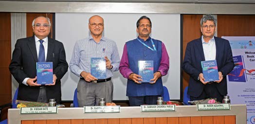


The diagnosis of parasitic diseases is the main research contributions in several areas related to parasitic serology and immunology; epidemiology of parasitic diseases of zoonotic importance and stool microscopy. Prof. Parija is known for his contribution in the development of simple, rapid and economical immunoassays for serodiagnosis and seroepidemiology of common parasitic diseases such as amoebiasis, hydatid disease, kala-azar, etc; and also newer methods in stool microscopy.
The diagnosis of parasitic diseases is the main field of interest and research. These include the modifications of existing immunoassays, and the development and evaluation of new immunoassays that are simple, sensitive, specific as well as inexpensive and reliable for their use in the diagnosis of common parasitic diseases in the poorly equipped laboratories.
Amoebiasis
Development of simple and rapid serological tests in the diagnosis of invasive amoebiasis and amoebic liver abscess, novel multiplex PCR to differentiate between pathogenic and non-pathogenic Entamoeba spp.
- Developed for the first time the carbon-immunoassay (CIA)and staphylococci adherence test (SAT) as two novel diagnostic tests for demonstration of amoebic antibodies in the serodiagnosis of amoebiasis. Since these two tests require only a compound light microscope and common reagents, the simplicity and economy of the methods would allow their adaptation at poorly equipped laboratories and doctor’s side clinic.
- Modified and simplified the IHA as a rapid –IHA for adaptation in the primary health centre level laboratories for diagnosis of amoebiasis.
- For the first time, the protein A- mediated haemagglutination (Protein A- IHA) was developed as a highly sensitive immunoassay for the diagnosis of amoebic liver abscess.
- Of particular importance is the detection of amoebic antigen in the serum obtained from the cases of amoebic liver abscess. The Co-A and CIEP were reported first by the author for detection of amoebic antigen in the serum. Both CIEP and Co-A tests were recommended as a simple and rapid slide agglutination tests for wide use in the field diagnosis of amoebiasis.
- Suggested the use of stool culture for amoeba as a diagnostic aid in the diagnosis of intestinal amoebiasis.
- For the first time documented Entamoeba dispar infections in humans in Pondicherry, India.
- For the first time documented Entamoeba moshkovskii human infection in India
- For the first time developed and evaluated multiplex PCR for simultaneous detection of Entamoeba histolytica, Entamoeba disparand Entamoeba moshkovskii in stool
- For the first time demonstrated the presence of Entamoeba histolytica DNA in the urine of patients for diagnosis of cases of amoebic liver abscesses.
- Demonstrated the existence of high degree of genetic variation exists among the clinical isolates of disparin Puducherry.
Cysticercosis
- For the first time documented and reported the incidence of ocular cysticercosis from Nepal.
- For the first time modified the indirect haemagglutination( IHA) test as a rapid –IHA by using Cysticercusantigen-sensitized double aldehyde stabilized chick RBCs in the test, in the diagnosis of cysticercosis.
- Evaluated the Dot-ELISA for diagnosis of neurocysticercosis in Pondicherry.
- For the first time conducted sero-epidemiological study of neurocysticercosis in Pondicherry.
- For the first time, it was demonstrated that Cysticercusantigens are excreted in the urine of patients with neurocysticercosis.
- Evaluated for the first time the bacterial Co-agglutination (Co-A), ELISA and Western blot for the detection of Cysticercus antigen in the urine for the diagnosis of neurocysticercosis.
- For the first demonstrated presence of Cysticercusantibodies in tear specimens for diagnosis of ocular cysticercosis.
Hydatid disease
Several immunoassays have been developed by Prof Parija for the diagnosis of hydatid disease. These would help not only in the diagnosis of an increased number of cases but also for large scale screening of the population for hydatid disease.
- For the first time, it was demonstrated that hydatid antigens are excreted in the urine of patients with hydatid disease.
- Evaluated for the first time the bacterial Co-agglutination (Co-A) and Counter-current immunoelectrophoresis (CIEP) as two simple, rapid and economical tests for the detection of hydatid antigens in the urine, a specimen collected by non-invasive method would be immensely useful for the diagnosis of hydatid disease, in poorly equipped laboratories in the developing countries like India.
- Evaluated and standardized for the first time the bacterial Co-agglutination (Co-A) as a simple and rapid slide agglutination test and Counter-current immunoelectrophoresis (CIEP) as a specific immunoassay for diagnosis of hydatid disease by detection of circulating hydatid antigen in the serum.
- Demonstrated the value of serum hydatid antigen as a prognostic marker for the pre- and post-treatment evaluation of the cases of hydatid disease.
- For the first time modified the indirect haemagglutination (IHA) test as a rapid –IHA by using hydatid antigen-sensitised double aldehyde stabilized chick RBCs in the test, in the diagnosis of hydatid disease.
- Developed Protein A -IHA as a highly sensitive immunoassay for the diagnosis of hydatid disease.
- The combined use of intradermal Casonis skin test and IHA has been recommended for detection of a large number of cases with hydatid diseases.
- Evaluated for the first time the bacterial Co-agglutination (Co-A) and Counter-current immunoelectrophoresis (CIEP) to establish the hydatid aetiology of the suspected cyst by detection of hydatid antigen in aspirated cystic fluid.
- The authors reported for the first time the evaluation of lacto-phenol cotton blue (LPCB) as a temporary staining agent for the scolices and hooklets in the wet mount preparation of the hydatid fluid, as a new method of parasitic diagnosis of hydatid cyst.
- The cases of hydatid disease were first documented and reported by the author from Pondicherry and conducted sero-epidemiological studies in hydatid disease in Pondicherry.
Malaria
- For the first time documented and reported the genotype of drug resistance-associated genes pvmdr-1 and pvcrt-o from Pondicherry and Jodhpur.
- Studied strain-specific phenotypes and the mechanism of gene regulation involved in chloroquine drug resistance using the whole transcriptome sequence profiling.
- Reported global comparative transcriptome profiling (RNA-Seq) to characterize the difference in the transcriptome between 48-h intraerythrocytic stage of chloroquine-sensitive and chloroquine-resistant P. falciparum (3D7 and Dd2) strains.
- Suggested the key contribution of ‘Plasmodium falciparum chloroquine resistance transporter protein’ to chloroquine resistance using iIn silico modeling of and biochemical studies.
- Studied degree of genetic diversity of PvMSP3βgene among the isolates collected from various parts of India.
Blastocystis
- For the first time documented and reported the subtype identification of Blastocystis in Pondicherry
- Reported severe blastocystis subtype 3 infection in a patient with colorectal cancer.
- Documented various morphological and reproductive features of Blastocystisusing various staining techniques on cultures positive in Jones’ medium
Kala-azar
Epidemiology of kala-azar in Nepal is the noted contribution
- A study was conducted to know the knowledge, attitude and perceptions of rural people staying in two villages’ endemic for kala-azar in Nepal. This study which was the first of its kind in kala-azar, has shown the usefulness and importance of such studies in initiating effective control measures in a community.
- For the first time, the cases of imported cutaneous leishmaniasis have been reported by the author in Nepal.
- The cases of post-kala-azar dermal leishmaniasis (PKDL) in Nepal have been detected and reported for the first time by the author.
- A case of kala-azar presenting with ulcers on the skin of the leg, the most unusual feature of kala-azar in Nepal have been documented for the first time.
- A case of kala-azar presenting with ulcers on the skin of the leg, the most unusual feature of kala-azar in Nepal have been documented for the first time.
- An extensive and laboratory study of kala-azar in children has highlighted for the first time the seriousness of the problem of kala-azar in children eastern Nepal, endemic for kala-azar.
- The anti-kala-azar treatment schedule has been recommended in Nepal.
- For the first time documented and reported cases of kala-azar without hepatomegaly in Nepal
Stool microscopy
Newer methods of stool microscopy and concentration methods for detection of intestinal parasitic infections is the main field of interest.
- Suggested sucrose layer centrifugation as a better method for concentration of stool for parasitic cysts and ova.
- For the first time evaluated and reported the use of lacto-phenol cotton blue (LPCB) a stain widely used in mycology, as a new and excellent temporary staining procedure for wet mount preparation of stool for intestinal cysts and ova in the stool for the diagnosis of intestinal parasitic infections.
- For the first time showed the usefulness of LPCB stain in the wet mount examination of stool for intestinal coccidian parasites such as Cyclospora and Isospora.
- For the first time evaluated and reported the use of potassium hydroxide (KOH), a stain widely used in mycology as a newtemporary staining procedure for wet mount preparation of stool for intestinal cysts and ova in the stool for the diagnosis of intestinal parasitic infections.
- For the first time showed the usefulness of thick stool smear for increasing sensitivity of stool microscopy in the diagnosis of intestinal parasitic infections.
- Studied the distribution pattern of the intestinal parasites among rural and urban communities in Puducherry.
Intestinal coccidian infections
- For the first time documented Cryptosporidium, Cyclospora andIsosporainfections in humans in Pondicherry, India.
- For the first time showed the usefulness of LPCB stain in the wet mount examination of stool for intestinal coccidian parasites such as Cyclospora and Isospora.
- Determined the intestinal parasitic profile in immunocompromised patients and also assessed the diagnostic accuracy of the ICT using ImmunoCard STAT kit in detecting Cryptosporidium
Enterobiasis
Epidemiology and microscopic diagnosis of enterobiasis
- For the first time documented and reported the prevalence of enterobiasis in children from Pondicherry
- For the first time evaluated and developed LPCB for the demonstration of Enterobius vermicularis eggs in the anal scrapings collected by the Scotch cellophane tape method.
Acanthamoeba and Microsporidia
For the first time documented and reported Acanthamoeba and microsporidia from corneal scrapings using conventional PCR in Puducherry.
Filariasis
The rapid –IHA and Protein A –IHA have for the first time for use in the laboratory diagnosis of lymphatic filariasis.
Hookworms
- For the first time documented and reported the prevalence of Necatoramericanus and Ancylostoma duodenale infections in Pondicherry.
- Evaluated the utility of conventional polymerase chain reaction for detection and species differentiation in human hookworm infections.
Ascariasis
Suggested the value of ultrasonography in the diagnosis of intestinal ascariasis.
Bacteriology
- Studied rate of hVISA among the MRSA isolates using PAP-AUC method and the distribution of mobile genetic element that carries methicillin-resistance gene SCCmec(Staphylococcal cassette chromosome mec)
- Studied various types of β-lactamases among the carbapenem-resistant isolates and epidemiological typing of baumanniiby MLST from isolates collected across India.
- Studied the molecular profile and resistance patterns of ESBLs among clinical isolates of Escherichia coli and Klebsiella pneumoniae at four tertiary care centres in India.
Animal studies
- Studied the effect of meloxicam (NSAID) on toxico-pathological and hematological parameters in Wistar rats.
- Studied the potential of adipose-derived mesenchymal stem cell (AD-MSCs) to enhance the rate of healing of full-thickness excisional skin wounds in rabbits.
